Current Pediatric Research
International Journal of Pediatrics
Prevalence of Vitamin D deficiency in Saudi children and risks of iron deficiency anemia.
1Department of Pediatrics, King Abdulaziz Medical City, Western Region, Ministry of National Guard, Health Affairs, Saudi Arabia
2King Abdullah International Medical Research Center, King Saud bin Abdulaziz University for Health Sciences, Jeddah, Saudi Arabia
3Department of Medicine, King Abdulaziz University, Saudi Arabia.
4College of Medicine, Ibn Sina National College, Saudi Arabia.
- *Corresponding Author:
- Faris Alzahrani
King Abdullah International Medical Research Center
Prince Abdulmajeed District, 5103, Jeddah, Makkah, Saudi Arabia.
Tel: +966555272894
E-mail: faris.a.alzahrani@hotmail.com
Accepted date: March 07, 2017
Background: Vitamin D is a prohormone that starts to synthesize in the skin and then convert from inactive to active metabolite through liver and kidney. It is essential nutrient for bone mineralization and deficiency can lead to rickets in children and osteomalacia in adult. In this study, we aim to estimate the prevalence of vitamin D deficiency in Saudi children and whether it is associated with iron deficiency anemia in Saudi children. Method: A combined design was used for the study, where we initially collected data from patients' medical records, retrospectively. Then, we compared between patients with vitamin D deficiency and iron deficiency anemia. We used data collection sheet composed of three sections; demographic data, laboratory results and growth parameters. Data were entered and analyzed using IBM Statistical Package for Social Sciences (SPSS) version 20. Results: From 401 patients reviewed, only 249 were eligible to enter the study. Male patients accounted for the 59.4%. The mean age for the patients was 6.14 years. One hundred patients had vitamin D deficiency (40.2%); whereas 21 patients had iron deficiency anemia (8.4%). Our data showed that there was no association between vitamin D deficiency and iron deficiency anemia (p value=0.505). Conclusion: To the contrast of global and local results, the prevalence of vitamin D deficiency was low in our study. We could not demonstrate any significance of iron deficiency anemia as an independent risk factor for vitamin D deficiency.
Keywords
Vitamin D deficiency, Iron deficiency anemia, Saudi children.
Introduction
Vitamin D is a prohormone that starts to synthesize in the skin and then convert from inactive to active metabolite through liver and kidney. It is essential nutrient for bone mineralization and deficiency can lead to rickets in children and osteomalacia in adult [1,2]. American Academy of Pediatrics (AAP) recommends 400 IU/day in children less than 1 year of life and 600 IU/day for older children as normal daily requirement.
The prevalence of vitamin D in healthy Saudi men was estimated to be 87.8% [3]. In the Eastern Province of Saudi Arabia, vitamin D deficiency was estimated to be more than 65%, despite adequate exposure to sun and consumption of dairy products [4]. Anemia is estimated to affect nearly 1.62 billion people around the globe. Many conditions can lead to anemia; including chronic kidney diseases or nutritional deficiencies. However, iron deficiency anemia is considered to be the cause in almost half of the cases [5]. Although iron deficiency anemia affects mainly children, elderly and young women, it can occur in all age groups [5,6].
The association between vitamin D deficiency and iron deficiency anemia has been studies [7,8]. Many studies showed that vitamin D is independently associated with lower concentration of hemoglobin and subsequently iron deficiency anemia [9]. However, other studies showed that vitamin D deficiency is not a risk factor for iron deficiency anemia [10].
In this study, we aim to estimate the prevalence of vitamin D deficiency in Saudi children and whether it is associated with iron deficiency anemia in Saudi children attending King Abdulaziz Medical City, Ministry of National Guard–Health Affairs–Western Region. Furthermore, we aim to estimate the prevalence of iron deficiency anemia in children with vitamin D deficiency [11].
Materials and Methods
Study Population
Initially, a cross-sectional chart-review retrospective study was conducted between January 2014 and December 2015 to estimate prevalence of vitamin D deficiency of pediatric patients who visited general pediatrics clinics for various medical conditions; but we excluded patents with other causes of anemia or conditions that may contribute to low concentration of hemoglobin; such as, chronic kidney disease, congenital heart disease, congenital anomalies, neurological diseases, endocrine abnormalities, liver diseases and intestinal malabsorption. We also included patients who had, at the time of the visit, results of complete blood count and vitamin D concentrations. Then those patients who fulfilled inclusion criteria were evaluated in retrospective cohort study where patient were grouped according to status of iron deficiency concentrations.
Data Collection
Data collection sheet composed of 3 sections was used to retrieve relevant information from medical records department and QuadraMed System.
The first section was about demographic data; including: date of birth, age and sex.
Second section was about biochemical profiles of 25OH and hemoglobin. This section was divided into two parts. The first part included: (vitamin D concentrations and supplements). We considered the patients having vitamin D deficiency when vitamin D 25 OH concentrations are below 50 nmol/l. The second part included: (iron concentrations, hemoglobin concentrations, Mean Corpuscle Volume (MCV), Mean Hemoglobin Concentration (MCH), Red Cell Distribution Width (RDW)). Anemia was reported when hemoglobin concentrations are below 11 g/dl; with the utilization of other parameters as well. Serum ferritin, iron, transferrin receptors were not included in the study as it is not routinely ordered in our institution.
The third section contained information about growth. It included: growth by height, growth by weight and Body Mass Index (BMI).
Statistical Analysis
The collected data were analyzed using IBM SPSS version 20. Simple descriptive statistics was produced in forms of means and medians. Frequencies and percentages were used to summarize qualitative data. For comparative statistics, Chi-square test was utilized considering p value 0.05 being two-tailed.
Ethical Approval
As this was a retrospective study, no informed consent was warranted from parents or guardians of patients. This study was approved by Institutional Review Board (IRB), King Abdullah International Medical Research Center (KAIMRC), protocol number: RJ15/037/J.
Results
Population
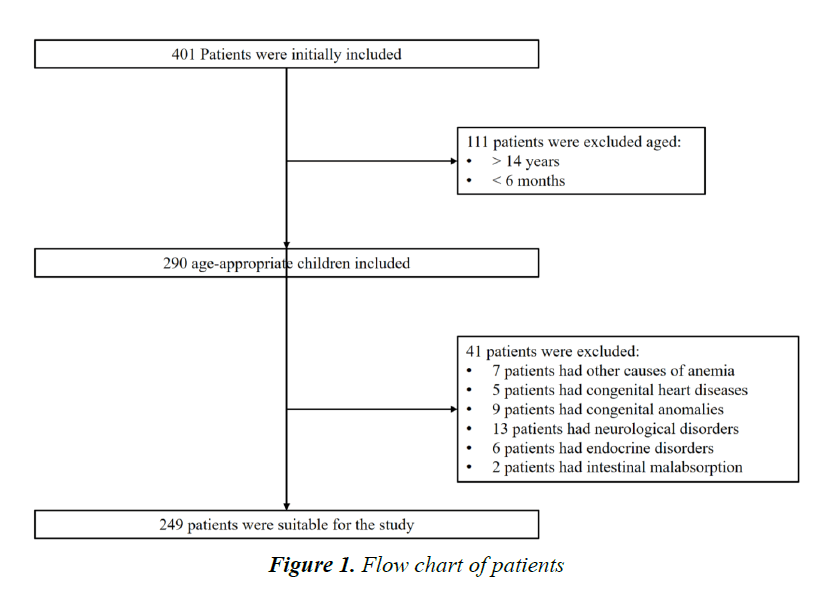
During the period of the study, 401 patients attended the general pediatrics clinic in King Abdulaziz Medical City– Western Region. A Total of 290 patients between the age of 6 months and 14 years were included initially (168 boys and 122 girls).
Out of those, 41 patients were exclude; 7 patients had other causes of anemia, such as, sickle cell anemia and Glucose- 6-phosphate dehydrogenase deficiency (G6PD deficiency), 5 patients had congenital heart diseases, 9 patients had congenital anomalies, 13 patients had neurological disorders, 6 patients had endocrine abnormalities, and 2 patients had intestinal malabsorption (Figure 1).
Only 249 were eligible to enter the study. Male patients accounted for the 59.4% and the females were 40.6%. The mean age for the patients was 6.58 years. The mean height for the patients was 111.20 cm, whereas the mean weight was 23.22 kg. The mean body mass index (BMI) was 17.17 kg/m2 (Table 1).
Laboratory Data
Out of the 249 patients, 100 had vitamin D deficiency (40.2%); whereas 21 patients had iron deficiency anemia (8.4%). The means levels vitamin and hemoglobin was 57.99 nmol/l and 12.47 g/dl, respectively. The mean value for MCV was 77.48 fl, whereas the mean value for MCH was 25.97 pg/cell, and the mean value for RDW was 14.4% (Table 2).
Utilizing Chi Square, there was no association between vitamin D deficiency and iron deficiency anemia (p value=0.505) (Table 3).
Furthermore, there was no difference between males and females in vitamin D deficiency (p value=0.090) or iron deficiency anemia (p value=0.102). Of the 100 patients with vitamin D deficiency, 50 children were taking vitamin D supplements. However, taking vitamin D supplements did not seem to affect vitamin D deficiency (p value=0.141) (Table 4, Table 5).
Discussion
Vitamin D Deficiency (VDD) is considered a common metabolic deficiency on Saudi Arabia. In the eastern province, VDD was estimated to be 65% (4); while among Saudi men, the prevalence was estimated to be 87% (3). To the contrast of these results, the prevalence in our institution was estimated to be at lower percentage compared to previous studies; the prevalence of vitamin D deficiency was 40.2%. This may be attributed to the increases awareness regarding vitamin D deficiency and the importance of supplements; as approximately 44.4% of our patients were taking vitamin D supplements.
| Characteristic | Mean (or Proportion) | 95% CI |
|---|---|---|
| Age (years) | 6.58 | 5.9442?6.8871 |
| Sex (males) | 59.4% | ? |
| Height (in cm) | 111.20 | ? |
| Weight (in kg) | 23.22 | ? |
| BMI (in kg/cm2) | 17.17 | 56.9353?67.4809 |
Table 1.General characteristics in population
| Variable | (Means ± SD) |
|---|---|
| Vitamin D Levels | 61.21 ( ± 28.85) |
| Hemoglobin Levels | 12.44 ( ± 1.12) |
| MCV | 78.01 ( ± 8.34) |
| MCH | 26.30 ( ± 4.61) |
| RDW | 14.46 ( ± 6.06) |
Table 2.Biochemical levels
| Iron Sufficient Patients | Iron Deficient Patients | P value | |
|---|---|---|---|
| Vitamin D levels (mean ± SD) | 57.36 (± 24.21) | 51.45 (± 25.29) | 0.446 |
| Vitamin D Deficiency (%) | 52 | 53.7 | 0.678 |
Table 3. Vitamin D comparison
| Variable | Vitamin D Sufficient | Vitamin D Deficient | P value |
|---|---|---|---|
| Age (mean ± SD) | 5.9 (± 3.71) | 8.2 (± 3.48) | 0.376 |
| Sex (males) | 60.62% | 55.36% | 0.48 |
| BMI (mean ± SD) | 17.23 (± 4.05) | 19.88 (± 10.73) | <0.0001 |
| Taking Supplements | 42.19% | 51.79% | 0.203 |
Table 4. Demographics and characteristics
| Variable | Vitamin D. Sufficient | Vitamin D. Deficient | P value |
|---|---|---|---|
| Hemoglobin Levels (mean ± SD) | 12.42 ( ± 1.21) | 12.55 ( ± 1.12) | 0.395 |
| MCV (mean ± SD) | 77.76 ( ± 8.13) | 76.54 ( ± 6.19) | 0.393 |
| MCH (mean ± SD) | 26.03 ( ± 2.43) | 25.97 ( ± 3.73) | 0.136 |
| RDW (mean ± SD) | 14.48 ( ± 7.26) | 14.13 ( ± 1.44) | 0.631 |
Note: Hemoglobin levels → g/dl; MCV → fl; MCH →pg; RDW →%; Vitamin D Ã ng/ml; Age → years; Height → cm; Weight → kg; BMI → kg/cm2.
Table 5. Biochemical levels.
There was no significant statistical difference among males and females in regard to the prevalence of vitamin D deficiency. Also, children with vitamin D deficiency were older with mean age of 8.2 years, in contrast to children without vitamin D deficiency; however, this, as well, was not statistically difference.
We noticed that children who were obese did have higher incidence of vitamin D deficiency compared to those with lower BMI (p value ≤ 0.0001). However, we could not identify such significance with iron deficiency anemia.
In contrast to previous studies (8–11), our results did not demonstrate any difference in regard to the prevalence of iron deficiency anemia in children with vitamin D deficiency; hence, we found no association between iron deficiency anemia and vitamin D deficiency.
The prevalence of iron deficiency anemia in our study was low compared to other studies [5,6].
Conclusion
To the contrast of global and local results, the prevalence of vitamin D deficiency was low in our study. Furthermore, iron deficiency anemia was not as prevalent as estimated internationally. However, due to the limited number of patients in our study, we think it will be difficult to generalize the results.
Acknowledgement
We greatly thank the Department of Medical Records, Mrs. Intesar Abdullah and Mr. Gazai.
References
- Misra M, Pacaud D, Petryk A, et al. Vitamin D deficiency in children and its management: review of current knowledge and recommendations. Pediatrics 2008; 122: 398-417.
- Perrine CG, Sharma AJ, Jefferds MED, et al. Adherence to vitamin D recommendations among US infants. Pediatrics 2010; 125: 627-632.
- Ardawi MSM, Sibiany AM, Bakhsh TM, et al. High prevalence of vitamin D deficiency among healthy Saudi Arabian men: Relationship to bone mineral density, parathyroid hormone, bone turnover markers, and lifestyle factors. Osteoporos Int 2012; 23: 675-686.
- Elsammak MY, Al-Wossaibi AA, Al-Howeish A, et al. High prevalence of vitamin D deficiency in the sunny Eastern region of Saudi Arabia: A hospital-based study. East Mediterr Health J 2011; 17: 317-322.
- Shams S, Asheri H, Kianmehr A, et al. The prevalence of iron deficiency anaemia in female medical students in Tehran. Singapore Med J 2010; 51: 116?119.
- Stevens GA, Finucane MM, De-Regil LM, et al. Global, regional and national trends in haemoglobin concentration and prevalence of total and severe anaemia in children and pregnant and non-pregnant women for 1995-2011: A systematic analysis of population-representative data. Lancet Glob Heal 2013; 1: e16?25.
- Golbahar J, Altayab D, Carreon E, et al. Association of vitamin D deficiency and hyperparathyroidism with anemia: A cross-sectional study. J Blood Med 2013; 4: 123?128.
- Yoon JH, Park CS, Seo JY, et al. Clinical characteristics and prevalence of vitamin D insufficiency in children less than two years of age. Korean J Pediatr 2011; 54: 298?303.
- Patel NM, Gutiérrez OM, Andress DL, et al. Vitamin D deficiency and anemia in early chronic kidney disease. Kidney Int 2010; 77: 715-720.
- Abdul-Razzak KK, Khoursheed AM, Altawalbeh SM, et al. Hb level in relation to vitamin D status in healthy infants and toddlers. Public Health Nutr 2012; 15: 1683-1687.
- Atkinson MA, Melamed ML, Kumar J, et al. Vitamin D, race and risk for anemia in children. J Pediatr 2014; 164: 153?158.
